Connect with us


The Premature Eye
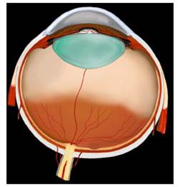
Premature eye with developed retina (blood vessels on orange-colored surface) and undeveloped retina (no blood vessels in peach- colored peripheral retina).
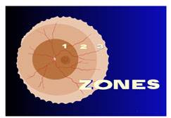
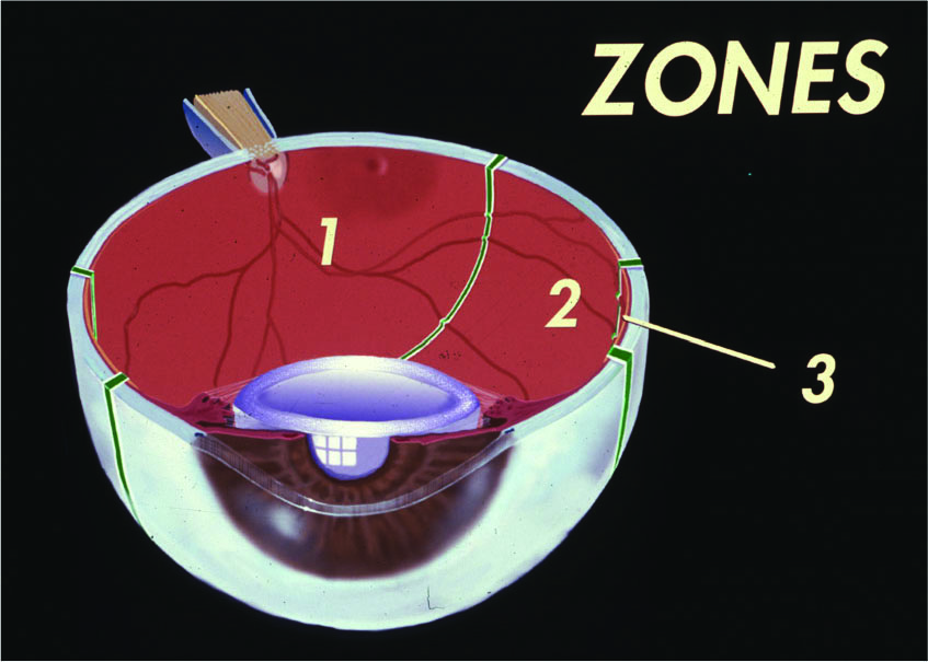
Two factors influence vision loss from ROP: the amount of retina that is undeveloped at the time of birth and the severity of the disease. The retina is divided into Zones 1, 2, and 3, and the severity of ROP is graded as Stage 1, 2, 3, 4, or 5.
The diagrams below show the zones of the premature retina. As the premature blood vessels grow from the back to the front of the eye, the amount of premature retina decreases. At full-term, the blood vessels extend to nearly the entire retina.